The Journal of Endodontics has published a new study on SWIFT MRI by which the authors compare several modalities of imaging, including cone beam CT, to the technique as it’s being developed. It looks very promising and is quickly becoming my favorite future technology in dentistry. The fact that it removes ionizing radiation from the equation really makes it a huge boon to dentistry all together.
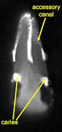
A resolution of 100 μm was obtained from in vitro teeth. SWIFT also identified the presence and extent of dental caries and fine structures of the teeth, including cracks and accessory canals, which are not visible with existing clinical radiography techniques. Intraoral positioning of the radiofrequency coil produced initial images of multiple adjacent teeth at a resolution of 400 μm.
100µm imaging is pretty amazing. Intraorally it looks like some more work will have to be done. 400µm really just doesn’t cut it. With technologies from Sirona where you will be able to merge Cerec restorations with cone beam studies at very high accuracy for predictable implant restorations, 400µm is just not detailed enough.